
Bladder TumourUro-Oncology Unit
Home » Uro-Oncology » Bladder Tumour
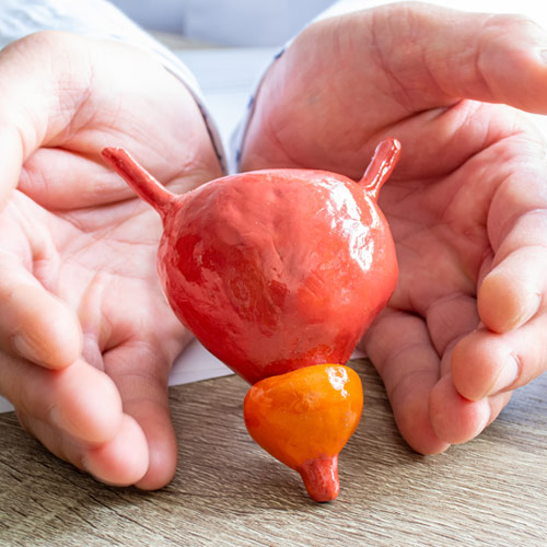
Bladder tumours develop from the transitional epithelium or urothelium that covers the inside of the bladder.
Bladder tumours are more frequent in men, and Spain has one of the highest incidences in Europe. Tobacco use is responsible for 50% of cases, increasing the risk by 4 in comparison to non-smoking patients. It also has a relationship with environmental and occupational exposure (aluminium, dyes, paints, oil, rubber, textiles, etc.). Other causes include radiation on the pelvis, parasites (schistosomiasis), drugs such as cyclophosphamide, exposure to trihalomethanes from ingested water etc.
The most frequent symptomatology is haematuria (80-90%), which may be accompanied by clots. Another form of presentation are irritative urinary symptoms (in the absence of urinary tract infection), consisting of increased urination frequency, dysuria or itching and urinary urgency.
Colic pain can also appear, as a consequence of upper urinary tract obstruction, or systemic symptoms in metastatic cases, being rare (weight loss, anorexia, fever etc).
There are two entities with different prognoses and treatments: the non-muscle invasive bladder tumour, which respects the muscle layer of the bladder (75%) and the muscle-invasive bladder tumour which affects the muscle layer (25%). Optimal management of this disease requires an uro-oncology unit capable of treating it in all its stages.
- Urologists highly specialised in bladder cancer.
- Ultrasound of the urinary system.
- Flexible urethrocystoscopy.
- Urine cytology.
- Uro-CT and thoracoabdominal-pelvic CT.
- IV Urography.
- BLADDER EPICHECK®
The ultrasound of the urinary system is fundamental to the diagnosis, and allows the evaluation of the presence of masses in the bladder, renal masses and obstruction of the upper urinary tract.
The cystoscopy is the best diagnostic test and is necessary to monitor patients with bladder tumours. It consists of an endoscopy of the urinary bladder using a cystoscope inserted through the urethra. This must be done under sterile conditions. It allows direct visualisation of bladder tumours, observing their number, location and appearance.
The urine cytology consists of the anatomopathological evaluation of the bladder cells that are released in the urine. This test helps predict the tumour grade and the presence of associated carcinoma in situ.
The Uro-CT is fundamental in the diagnosis and monitoring of patients with bladder tumours, since it allows the evaluation of ureters, pelvis and renal calyces to rule out the presence of tumour implants in these locations. Upper urinary tract tumours can coexist with bladder tumours in 2-4% of the cases, rising up to 7% if the bladder tumour is in the trigone. In addition, thoracoabdominal-pelvic CT is used to rule out the presence of metastasis.
- Transurethral resection of the bladder.
- Bladder biopsy.
- Intravesical instillations: mitomycin C, BCG.
- Radical cystectomy (by open, laparoscopic/robotic surgery).
- Bladder preservation (trimodal treatment: surgery, radiotherapy and chemotherapy).
- Close collaboration with the Medical Oncology and Radiotherapy services.
- Hypogastric artery embolisation (interventional radiology).
- Bladder formolisation.
Non-muscle invasive bladder tumour: treatment and monitoring.
The early treatment of the bladder tumour is the transurethral resection of the bladder (TUR). Consists of the exploration and removal of bladder lesions through endoscopy, curing non-muscle invasive bladder tumours. In addition, it allows obtaining enough tumour sample for the anatomopathological diagnosis of tumours affecting the muscle layer.
Sometimes it is necessary to administer intravesical treatments after TUR (mitomycin C, BCG) with the aim of preventing the recurrence and progression of these tumours. Several factors are taken into account to determine whether the patient needs these therapies: local stage, tumour grade, size of the lesions removed, history of previous recurrences, or presence of associated carcinoma in situ.
Patients with high-grade tumours, with poor prognostic factors and that recur or progress despite conventional treatments, are offered an early cystectomy, thus improving their long-term prognosis.
Monitoring is performed according to the risk of progression and is based on the periodic performance of cystoscopies or ultrasounds, urine cytologies and Uro-CTs.
Muscle-invasive bladder tumour: treatment and monitoring.
The first manoeuvre to be carried out is the TUR of the bladder, to establish the diagnosis of tumour infiltration of the muscle layer.
Once the diagnosis of a muscle-invasive bladder tumour has been made, the treatment of choice with curative intent is the radical cystectomy. It is a complex surgery that consists of the exeresis of the bladder, prostate and seminal vesicles in men, and the uterus, ovaries, cervix and front wall of the vagina in women. In addition, lymph nodes in the pelvis should be removed. During the same surgical procedure it is necessary to reconstruct the urinary tract so that the patient can urinate. There are various techniques for this purpose. The most commonly used is the neobladder (a new bladder is crafted out of the patient’s intestine), opening the ureters to the skin (cutaneous ureterostomy) or anastomosing the ureters to a portion of the intestine, which will act as a stoma (cutaneous ureteroileostomy). This surgery can be performed through an open, laparoscopic or robotic approach.
» Trimodal or multimodal therapy:
In very selected patients with tumours with favourable conditions, or in those patients who are not suitable for cystectomy, it is possible to preserve the bladder by subjecting the patient to a combined treatment. TUR of the bladder, chemotherapy and radiotherapy are offered.
» Chemotherapy based on regimens that include cisplatin.
It should be offered before cystectomy (neoadjuvant regimen) in patients without nodal or visceral metastases, achieving improvements in survival of up to 8% in 5 years. After cystectomy, it should be administered in patients with tumour involvement of perivesical fat or neighbouring organs, lymph node involvement, or in patients with metastases at diagnosis, who have not yet been operated. Immunotherapy is used as a second line of treatment once the disease progresses in spite of chemotherapy.
» Radiotherapy:
Used as part of the multimodal treatment or as mitigation therapy of The use of the drug is not a substitute for the use of the drug itself, but is a part of multimodal treatment or palliative treatment of intractable haematuria. Other palliative measures to combat haematuria in patients with incurable tumours include the embolisation of the hypogastric arteries or bladder formolisation.
These patients should be closely monitored by the urologist and the oncologist to prevent the progression of the disease.
