DiagnosisProstate Cancer
Home » Prostate Cancer » Diagnosis of prostate cancer
Diagnosis of prostate cancer
Screening for prostate cancer is first performed through a digital exam (digital rectal examination), and shall be complemented with a blood test to measure the PSA. It is possible to detect the disease before suffering the symptoms. Today, there is new diagnostic tests available that facilitate this task: proPSA, 4-Kallikrein, PCA3.
After a general physical exam, your doctor will ask you some questions about your symptoms and medical background, and will perform some of the following tests:
» Digital rectal exam (digital rectal examination):
During the test the doctor inserts a finger into the patient’s rectum to detect any hard, irregular areas in the prostate (swelling or protuberance) which could indicate cancer.
» Blood test:
Consists in determining the blood levels of prostate-specific antigen (PSA), a protein produced by the prostate and that can be raised in a number of situations, including PCa.
» Urine test:
A urine sample is taken to determine whether there is blood or other abnormalities, such as bacteria or inflammatory cells that could lead to an infection. It is also necessary to determine some cancer markers.

» Transrectal ultrasound (TRUS):
This technique provides a more accurate image of the prostate than traditional ultrasound. An ultrasound probe is introduced in the patient’s rectum. It is performed on an outpatient basis and lasts about 10 minutes.
It enables evaluating the shape and size of the prostate. It is also helpful while collecting biopsies. Performance for tumour detection through imaging is low.
» Prostate biopsy:
The only way to confirm the diagnosis of prostate cancer is by taking a tissue sample (biopsy).
The biopsy consists of inserting a needle into the prostate with the intention of extracting some of this tissue and analysing it. This test allows confirmation of the disease, although a negative result does not completely rule it out.
Fusion prostate biopsy
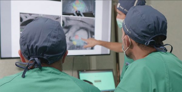
Given the scarce information offered by the transrectal ultrasound, nowadays it has been established that a multiparametric magnetic resonance shall be carried out before the first biopsy, which will show whether there are any suspected PCa areas.
With the MR image in the operating room, thanks to a complex computer system, the images of the MR are merged with those obtained at that time with the ultrasound, allowing us to direct the biopsies to the suspicious areas of the prostate.
The information obtained is much more comprehensive and will allow a personalised treatment of the patient (see specific section for more information).
